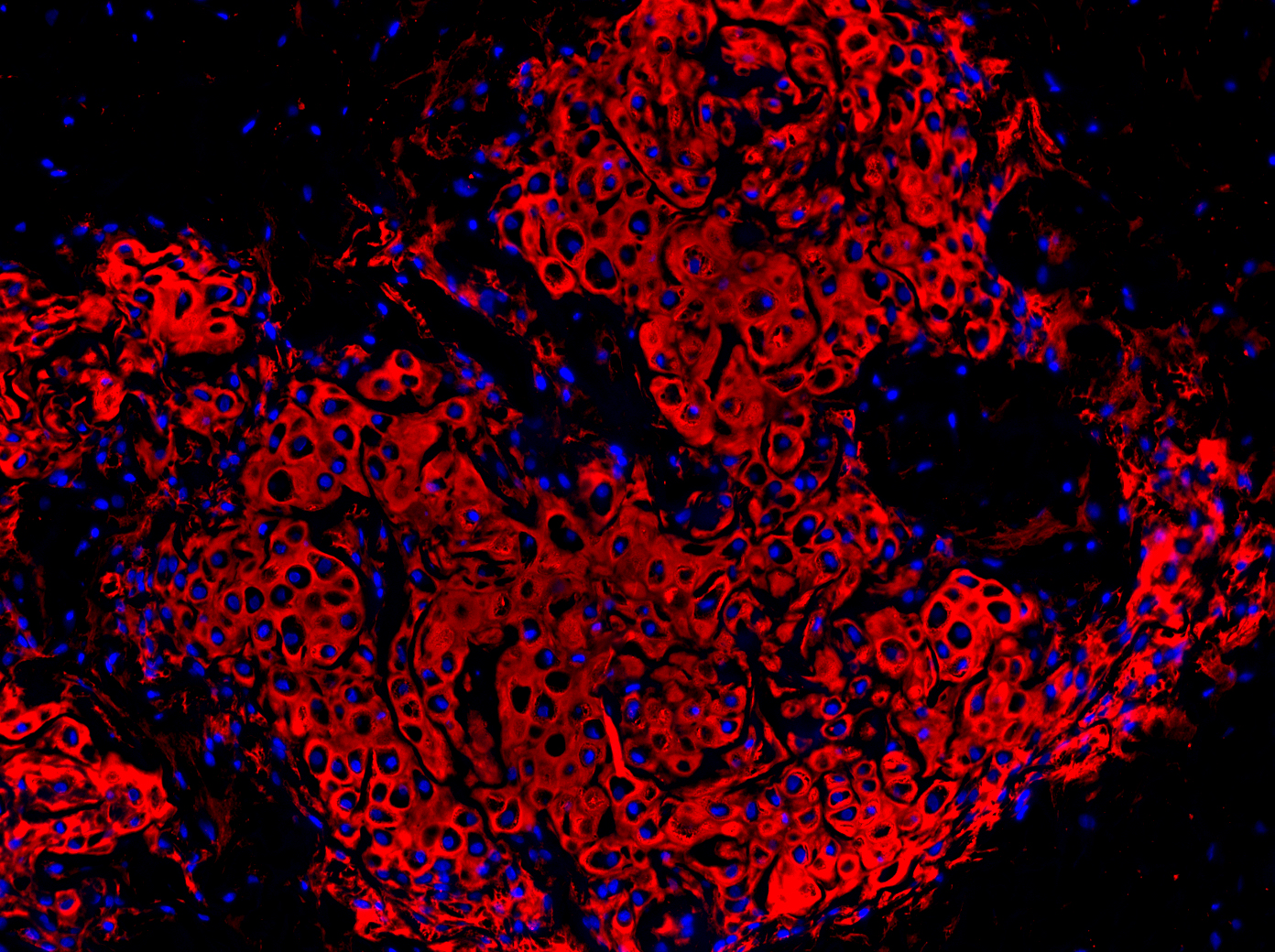
The cells reside within the temporomandibular joint (TMJ), which articulates the jaw bone to the skull. When the stem cells were manipulated in animals with TMJ degeneration, the cells repaired cartilage in the joint. A single cell transplanted in a mouse spontaneously generated cartilage and bone and even began to form a bone marrow niche.
The findings were published on Oct. 10 in Nature Communications.
«This is very exciting for the field because patients who have problems with their jaws and TMJs are very limited in terms of clinical treatments available," said Mildred C. Embree, DMD, PhD, assistant professor of dental medicine at Columbia and the lead author of the study. Dr. Embree’s team, the TMJ Biology and Regenerative Medicine Lab, conducted the research with colleagues including Jeremy Mao, DDS, PhD, the Edwin S. Robinson Professor of Dentistry (in Orthopedic Surgery) at Columbia.
Up to 10 million people in the United States, primarily women, have TMJ disorders, according to the National Institutes of Health. Options for treatment currently include either surgery or palliative care, which addresses symptoms but can’t regenerate the damaged tissue. Dr. Embree’s findings suggest that stem cells already present in the joint could be manipulated to repair it.
Cartilage helps to cushion the joints and allows them to move smoothly. The type of cartilage within the TMJ is fibrocartilage, which is also found in the knee meniscus and in the discs between the vertebrae. Because fibrocartilage cannot regrow or heal, injury or disease that damages this tissue can lead to permanent disability.
Medical researchers have been working to use stem cells, immature cells that can develop into various types of tissue, to regenerate cartilage. Given the challenges of transplanting donor stem cells, such as the possibility of rejection by the recipient, researchers are especially interested in finding ways to use stem cells already living in the body.
«The implications of these findings are broad," said Dr. Mao, «including for clinical therapies. They suggest that molecular signals that govern stem cells may have therapeutic applications for cartilage and bone regeneration. Cartilage and certain bone defects are notoriously difficult to heal." Dr. Mao is
In a series of experiments described in the new report, Dr. Embree, Dr. Mao, and their colleagues isolated fibrocartilage stem cells (FCSCs) from the joint and showed that the cells can form cartilage and bone, both in the laboratory and when implanted into animals. «I didn’t have to add any reagents to the cells," Dr. Embree said. «They were programmed to do this." And while some approaches to regenerating injured tissue require growth factors or biomaterials for the cells to grow on, she noted, the FCSCs grew and matured spontaneously.
Dr. Embree and her team also identified a molecular signal, Wnt, that depletes FCSCs and causes cartilage degeneration. Injecting a
She and her colleagues are now searching for other small molecules that could be used to inhibit Wnt and promote FCSC growth. The idea, according to Dr. Embree, will be to find a drug with minimal side effects that could be injected right into the joint.
Children with juvenile idiopathic arthritis can have stunted jaw growth that can’t be treated with existing drugs, Dr. Embree noted. Since the TMJ is a growth center for the jaw, the new research may offer strategies for treating these children and lead to a better understanding of how the jaw grows and develops. While orthodontists currently rely on clunky technologies like headgear to modify jaw growth, she added, the findings could point toward ways to modulate growth on the cellular level.
Ultimately, Dr. Embree and her team say, the findings could lead to strategies for repairing fibrocartilage in other joints, including the knees and vertebral discs. «Those types of cartilage have different cellular constituents, so we would have to really investigate the molecular underpinnings regarding how these cells are regulated," the researcher said.


