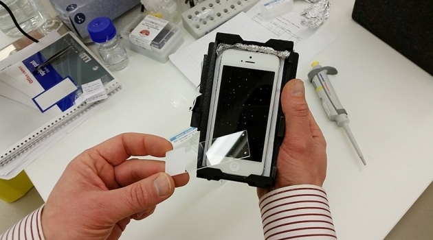
A small, simple and relatively cheap microscope which is printed using a 3D printer and coupled to the camera of a mobile phone can be used to assess tumours, bacteria, viruses and fungal cells. The microscope and mobile phone can detect the presence of a targeted DNA sequence, letting doctors know exactly which form of cancer, bacteria or virus they are dealing with. This type of analysis is called targeted DNA sequencing and is important for finding out which treatment would be most effective.
«I’m used to needing large, expensive and complicated machines that take up entire rooms to do DNA sequencing. To me, the thought of being able to use a mobile phone is very interesting. It opens the door to many new and very important areas of application," says Mats Nilsson at Stockholm and Uppsala University, and SciLifeLab Stockholm.
Today, DNA sequencing is done at the laboratories of large hospitals. The small, 3D printed microscope can make the technology available to more people, even in poorer parts of the world. If the microscope were to be manufactured on a larger scale, the price could drop below 500 dollars, and it is not dependent on a stable supply of electricity since it runs off the phone’s battery.
«Antibiotics are an efficient weapon against bacteria. But we are beginning to loose that weapon as bacteria become resistant. With tuberculosis, resistance is a serious problem. But if we could look at the DNA and find out if a bacteria is sensitive to a certain type of antibiotic, we could pick the right treatment straight away. That is where I believe this concept has its greatest potential," says Mats Nilsson.
In a new study published in Nature Communications, the technology has been used to diagnose colorectal tumours. By studying certain mutations of the tumour it is possible to dismiss certain treatments that are known not to work, and it also becomes possible to send pictures and information about the DNA to doctors elsewhere in the world. The researchers also hope the technology can speed up diagnosis of virus infections such as Ebola and Zika.
The microscope is a team effort between researchers at the California Nanosystems Institute of UCLA, who have built the actual microscope, and researchers at Stockholm and Uppsala University and SciLifeLab Stockholm, who have developed the DNA analysis.
Source: http://www.uu.se/en/media/news/article/?id=8064&area=2,4,10,16,24&typ=artikel&lang=en


