The
Images of the protein, which revealed several unusual features, are published in the Aug. 26 issue of the journal Science by structural biologist Filippo Mancia, PhD, assistant professor of physiology & cellular biophysics, who led a team of scientists including Wayne Hendrickson, Larry Shapiro, Joachim Frank, and Bill Blaner at Columbia University Medical Center, Loredana Quadro at Rutgers University, Chiara Manzini at George Washington University and David Weber at the University of Maryland School of Medicine.
New camera allows researchers to see smaller and smaller cellular components
Until the new study, the way STRA6 transports vitamin A into the cell had been a mystery. Most transporters interact directly with the substances they transport. But STRA6 only interacts with vitamin A via an intermediary protein that carries the greasy vitamin A in the bloodstream. Revealing the structure of STRA6 may not only give the researchers insight into vitamin A transport, but also clues about how other related transporters may work.
A new type of camera technology was a key element to getting the images of STRA6. When paired with an electron microscope, the camera allows biologists to see tiny,
«We can now get near atomic resolution because the new camera is much faster and allows us to take a movie of the molecules," says Oliver Clarke, PhD, an associate research scientist in the Hendrickson lab at Columbia University Medical Center. «Even under the electron microscope, the molecules are moving around by a tiny amount, but when you take a picture of something moving, it comes out blurry. With such a movie, we can align the frames of the movie to generate a sharper image.»
Imaging the molecule also depended on painstaking biochemical procedures, developed by Yunting Chen, PhD, an associate research scientist in the Mancia lab, to generate large quantities of the protein and separate them from the cell’s other components. «It’s a very delicate protein, and we had to mimic its environment to keep it from getting out of shape," she says. Those efforts took about two years to perfect.
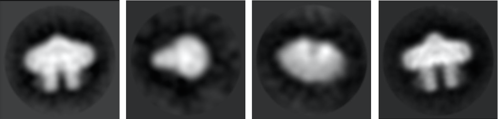
The researchers used approximately 70,000 individual pictures of STRA6 to generate a
Images reveal «unusual» features of vitamin A transport
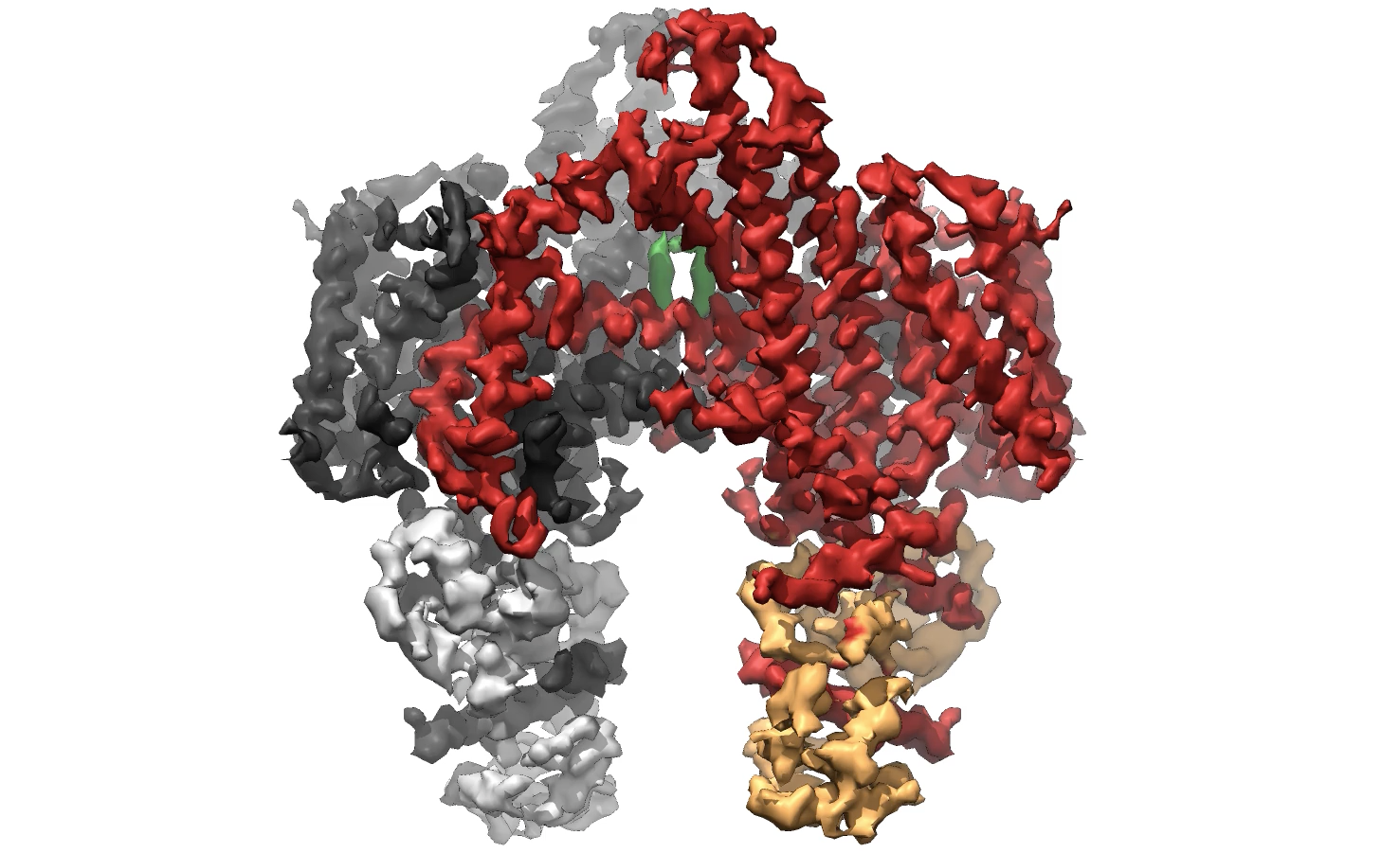
The images and model reveal STRA6 is «a bit of a freak," says Dr. Clarke. Even more surprising was the fact that STRA6 does not work alone but is instead tightly associated with another protein, calmodulin, which plays a key role in calcium signaling.
Although vitamin A moves through STRA6 to enter the cell, there is no channel in STRA6 like most transporters. Instead, vitamin A enters the top of STRA6 but then appears poised to exit through a side window that opens directly into the cell membrane, not the cell interior.
Though this needs to be verified, the mechanism may be a way to protect cells from too much vitamin A. «Vitamin A is actually somewhat toxic," says Dr. Mancia. «Trapping vitamin A inside the membrane may keep control of the amount inside the cell.»
The new model of STRA6 advances the understanding of a critical cellular function and may help researchers understand how other, still mysterious cellular components work.