
Developers
T.Boland,
Description of the technology
There are several approaches for obtaining organs for transplantation, and bioprinting is the most promising of them.
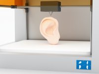
Bioprinting, or 3D bioprinting — is a technology intending to construct tissues and organs from cell aggregations; it is similar to a sort of constructor. Such an assembly, or bioprinting, is carried out on the specially developed 3D bioprinters, similar to that how various details are printed on 3D printers —
As the first printers for bioprinting, common table
Only patient’s own cells are used in bioprinting. Those may be monoculture of cells with terminal function (for example, cells of inner coat of vessels) or suspension of pluripotent stem cells which are able to form any tissue.

The first stage of bioprinting is the process of obtaining donor cells. They may be taken from virtually any tissue (for example, from the fat tissue). Afterwards, they are induced to differentiate in the required direction.
The second stage is the process of obtaining tissue sheroids. Each sheroid contains 10 to 20 thousands of cells and has a contact with many neighbouring speroids in the organ being constructed. That allows to achieve the high density of the cell structure, and that is a key factor determining quality of the printed organ and its functionality. Alternative variants of bioprinting use scaffolds — temporary supporting structures which hold cells. It is supposed that scaffolds have to be gradually destroyed while cells are synthetizing their own matrix; however, synchronization of those processes is hardly accessible. Moreover, scaffolds don’t give appropriate cell density.
On the third stage, the process of bioprinting proper takes place. Bioprinting is substantially similar to
Up to date, skin, cartilage, fragments of liver and kidney tissues and the model of thyroid gland were obtained using the technology of bioprinting.
Practical application
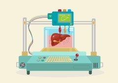
Scientists and physicians intend using 3D bioprinting in regenerative medicine. This technology is suitable for supplying the demand in tissues and organs for transplantation. Bioprinting has other applications as well. The main of them is toxicological studies of various compounds and new pharmaceuticals without use of laboratory animals. Another application is modeling of pathological processes for the study of key mechanisms of their pathogenesis.
Laboratories
- Organovo, San Diego, California (USA)
- RegenHU,
Villaz-St-Pierre (Switzerland) - 3D Bioprinting Solutions, Москва (Russia)
- Cyfuse Biomedical, Hongo,
Bunkyo-ku , Tokyo (Japan) - ROKIT,
Geumcheon-gu , Seoul (South Korea)
Links
make-3d .ru/articles/biopechat-organov-na-3d-printere /- http://www.computerra.ru/61217/
3d-bioprinting / - http://www.computerra.ru/135772/3d-bioprinting-
of-thyroid-gland / - https://www.google.com.ua/url?sa=t&rct=j&q=&esrc=s&source=web&cd=2&cad=rja&uact=8&ved=
0ahUKEwiZya-vmcDLAhWyZpoKHb5kCboQFggiMAE &url=https%3A%2 °F %2Fen.wikipedia.org%2Fwiki%2F3D_bioprinting&usg=AFQjCNH1uGM9kPSpxjREoSdeDzi2aUhg0w&sig2=0AcuirjsMxOOk3G4xnkJhg&bvm=bv.116636494,d.bGs - http://www.nkj.ru/archive/articles/23328/
Publications
- Mironov, Vladimir, Nuno Reis, and Brian Derby. «Review: bioprinting: a beginning." Tissue engineering 12.4 (2006): 631–634.
- Derby, Brian. «Bioprinting: inkjet printing proteins and hybrid
cell-containing materials and structures." J. Mater. Chem. 18.47 (2008): 5717–5721. - Murphy, Sean
v. , and Anthony Atala. «3D bioprinting of tissues and organs." Nature biotechnology 32.8 (2014): 773–785. - Cui, Xiaofeng, et al. «Direct human cartilage repair using
three-dimensional bioprinting technology." Tissue Engineering Part A 18.11–12 (2012): 1304–1312. - Norotte, Cyrille, et al. «
Scaffold-free vascular tissue engineering using bioprinting." Biomaterials 30.30 (2009): 5910–5917. - Kolesky, David B., et al. «3D bioprinting of vascularized, heterogeneous cell‐laden tissue constructs." Advanced materials 26.19 (2014): 3124–3130.