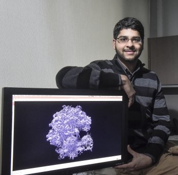
PhD candidate Ali Punjani helped develop a set of algorithms that can generate 3D structures of tiny protein molecules. The hope is that the discovery may revolutionize the development of drug therapies for a range of diseases. (Photo by Ken Jones)
«Designing successful drugs is like solving a puzzle," says U of T PhD student Ali Punjani, who helped develop the machine learning algorithms.
«Without knowing the
Watch the video explaining how this research works.
The ability to determine the 3D atomic structure of protein molecules is critical in understanding how they work and how they will respond to drug therapies, notes Punjani.
Drugs work by binding to a specific protein molecule and changing its 3D shape, altering the way it works once inside the body. The ideal drug is designed in a shape that will only bind to a specific protein or proteins involved in a disease while eliminating side effects that occur when drugs bind to other proteins in the body.
This new set of algorithms reconstructs 3D structures of protein molecules using microscopic images. Since proteins are tiny — even smaller than a wavelength of light — they can’t be seen directly without using sophisticated techniques like electron cryomicroscopy (
«Our approach solves some of the major problems in terms of speed and number of structures you can determine," says Professor David Fleet, chair of the Computer and Mathematical Sciences Department at U of T Scarborough and Punjani’s PhD supervisor.
The algorithms, which were
«Existing techniques take several days or even weeks to generate a 3D structure on a cluster of computers," says Brubaker. «Our approach can make it possible in minutes on a single computer.»
Punjani adds that existing techniques often generate incorrect structures unless the user provides an accurate guess of the molecule being studied. What’s novel about their approach is that it eliminates the need for prior knowledge about the protein molecule being studied.
«We hope this will allow discoveries to happen at a
The research, which included a collaboration with U of T Professor John Rubinstein, a Canada Research Chair in Electron Cryomicroscopy, received funding from the Natural Sciences and Engineering Research Council of Canada (NSERC). It’s also been published in the current edition of the Journal Nature Methods.
Meanwhile, the team’s


